



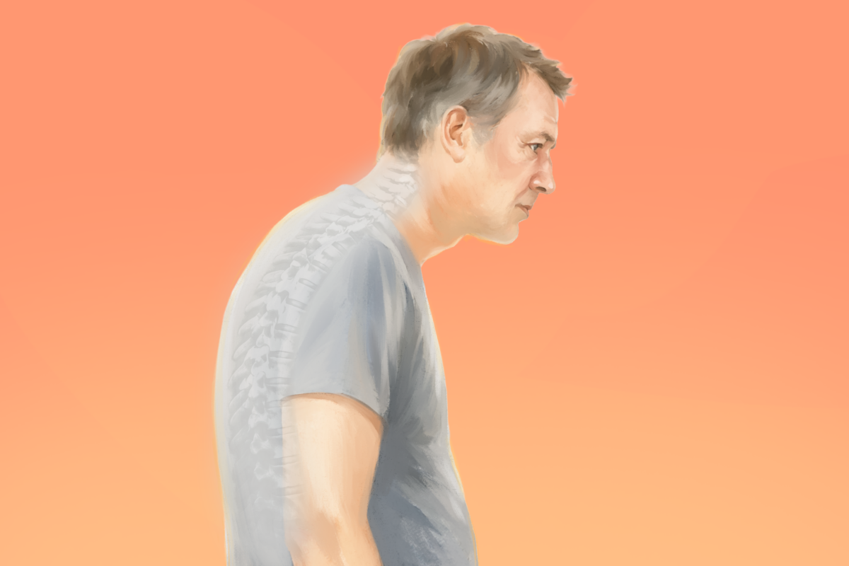
Many people know it as a “hunchback” or “dowager’s hump,” but doctors use a different term: hyperkyphosis.
Confusingly, it’s often shortened to simply kyphosis—even though kyphosis actually refers to the normal, gentle forward curve of the upper spine that everyone has.
That natural curve helps you stay balanced, absorb shock, and stand upright. It’s only when the curve becomes excessively pronounced that it becomes problematic.
-
A visibly rounded upper back, the hallmark and most noticeable sign
-
Mild to severe back pain, often worsened by movement
-
A forward-leaning head posture
-
Uneven shoulder height
-
Tight hamstrings
-
Fatigue
In more severe cases, symptoms can include:
-
Chest, back, or neck pain as surrounding muscles and joints compensate for the altered posture
-
Shortness of breath
-
Muscle weakness
-
Numbness or tingling
[shortcut_anchor id=”anchor_1785566863850″ label=”Causes”]What Are the Causes of Kyphosis?[/shortcut_anchor]The spine consists of 24 vertebrae, or small bones stacked together to form the spinal column and protect the spinal cord. Intervertebral discs absorb shock and reduce stress between the bones.
Viewed from the side, a healthy spine has three natural curves: two inward curves at your neck and lower back called cervical and lumber spines, and one outward curve at your chest called the thoracic spine.
Some kyphosis is caused by your posture, and those are reversible, while other types may not be.
1. Postural Kyphosis
This is the most common type of kyphosis and typically develops during adolescence, more often in girls. It is caused by habitual slouching or weak back muscles, not by any abnormality in the vertebrae themselves.
When you slouch, the muscles in your upper back become chronically lengthened and may weaken, while the chest muscles shorten and tighten. This muscle imbalance encourages a rounded-shoulder posture and makes it more difficult to maintain an upright position.
However, your spine curve is flexible. As long as you change how you sit and stand, over time you can train your muscles back. Postural kyphosis is usually painless and rarely progresses. Some people may compensate with an exaggerated inward curve of the lower back.
2. Scheuermann’s Kyphosis
This is a structural form of kyphosis that also typically develops during adolescence but is more common in boys. The curve usually measures 45 to 75 degrees and generally stops progressing once bone growth is complete.
Unlike postural kyphosis, the spinal curve is rigid and cannot be corrected by standing up straight. It is caused by abnormal vertebral growth, in which three or more consecutive vertebrae grow into a wedge shape, creating a sharp, angular curve, most often in the upper back.
X-rays typically show wedge-shaped vertebrae, narrowed disc spaces, and sometimes small herniations called Schmorl’s nodes.
The exact cause of this type of kyphosis is unknown. One early theory, proposed by Danish radiologist Holger Scheuermann, after whom this condition is named, suggested that reduced blood flow to the cartilage in growing vertebrae disrupted normal bone development. Currently, most researchers believe that it begins with damage to the vertebral growth plates during adolescence. Uneven bone growth causes the front of the vertebrae to grow slower than the back, resulting in wedge-shaped bones that can contribute to kyphosis. Other likely contributing factors may include how the spine is structured, moves, and responds to forces during activities, as well as mild osteoporosis, muscle abnormalities, and genetics.
3. Congenital Kyphosis
This type of kyphosis is present at birth and results from abnormal spinal development in the womb, such as vertebrae that didn’t fully form or were fused together. The spinal curvature typically worsens as the child grows and often requires early surgery to prevent progression. Congenital kyphosis is frequently associated with other birth defects. In severe cases, the condition can lead to neurological complications.
Kyphosis can also result from many medical conditions, such as:
-
Degenerative Kyphosis: This is the most common type in older adults. Age-related wear, such as disc degeneration, arthritis, and osteoporosis, causes discs to collapse and ligaments to weaken, gradually pulling the spine out of alignment. As alignment worsens, mechanical stress increases, which can accelerate further degeneration. This type of kyphosis often comes with back pain and stiffness.
-
Traumatic Kyphosis: This type develops as a result of spinal injury, such as vertebral compression fractures, ligament damage, or other trauma affecting spinal stability. Fractures in the mid or lower back commonly cause some degree of kyphotic deformity and may progress or cause nerve-related complications.
-
Neuromuscular (Paralytic) Kyphosis: This type occurs when conditions that cause muscle weakness or paralysis, such as cerebral palsy, muscular dystrophy, polio, spinal muscular atrophy, and spina bifida, reduce the spine’s ability to hold proper alignment. Unlike sudden injuries, neuromuscular kyphosis develops slowly as muscle weakness and imbalance build up over time.
-
Tumor-Related Kyphosis: This type is caused by tumors that weaken, damage, or destroy the vertebrae—most often cancer that has spread to the spine, but also primary bone cancer or multiple myeloma (a blood cancer originating in bone marrow). Chemotherapy or radiation to the spine can also increase the risk of vertebral compression fractures and wedge-shaped vertebrae.
-
Spondylolisthesis-Associated Kyphosis: Spondylolisthesis is a condition in which one vertebra slips forward relative to the vertebra below it. This first tends to increase the inward curve of the lower back (lordosis) and, in more advanced cases, can contribute to kyphosis as the spine loses overall balance.
-
Infectious (Post-Infectious) Kyphosis: This type results from infection that destroys vertebral bone, most often in the upper or mid back. It is usually a long-term complication of a severe spinal infection, such as tuberculosis or bacterial bone infection (osteomyelitis).
Risk Factors
In addition to age (the biggest overall risk factor) and family history, common risk factors include:
-
Smoking: Smoking may increase the risk of developing kyphosis by contributing to decreased bone density, spinal degeneration, and a higher likelihood of vertebral compression fractures.
-
Sedentary Lifestyle: Lack of physical activity and weak back muscles can contribute to postural kyphosis by affecting spinal alignment and support.
-
Low Bone Mineral Density: Weakened bones are more prone to vertebral compression fractures, which can contribute to kyphotic curvature.
[shortcut_anchor id=”anchor_1785566915939″ label=”Diagnosis”]How Is Kyphosis Diagnosed?[/shortcut_anchor]Although a rounded or hunched back may be noticeable, kyphosis—especially the non-postural or severe type—requires a medical diagnosis to confirm that the spinal curvature is abnormal and to determine its cause and severity. Diagnosis also guides treatment decisions.
The evaluation will typically include three steps:
1. Medical History
A doctor reviews past spinal X-rays, observes posture and movement, and asks about the onset of the symptoms or spinal rounding, changes in height, family history, pain, neurological symptoms, such as numbness, tingling, muscle weakness, or bowel and bladder dysfunction, and any previous spinal injuries or surgeries.
2. Physical Examination
The healthcare provider checks the shape and flexibility of the spine by observing posture, examining the back, and evaluating range of motion. They also check for tenderness, muscle strength, sensation, reflexes, and signs of nerve involvement. A key part of the examination is determining whether the spinal curve is flexible or rigid.
3. Additional Tests
The doctor may order several tests, including:
-
X-Rays: Uses a standing lateral (side-view) full-spine X-ray, with a spinal curve greater than 50 degrees generally considered abnormal.
-
Computed Tomography (CT) Scan: Uses X-rays to create detailed cross-sectional and three-dimensional images of the spine. It provides more detailed information than a standard X-ray and is particularly useful for evaluating spinal fractures and bone abnormalities.
-
Magnetic Resonance Imaging (MRI): Uses magnetic fields and radio waves to produce detailed images of the spine, including the discs, nerves, spinal cord, and surrounding soft tissues. It is used to evaluate nerve or spinal cord compression that may be causing neurological symptoms, such as pain, weakness, numbness, or impaired movement. In some cases, a contrast agent may be used to improve visualization of specific tissues.
-
Bone Scintigraphy (Bone Scan): Detects areas of unusual bone activity, such as inflammation, infection, fractures, or other abnormalities affecting the spine.
-
Electromyography and Nerve Conduction Studies: Measure the electrical activity of the muscles and nerves to help detect nerve damage, nerve compression, or disorders affecting nerve and muscle function.
-
Pulmonary Function Tests: If the spinal curve is severe, pulmonary function tests may be performed to determine whether kyphosis has reduced chest space and restricted lung function or breathing.
-
Biopsy: Involves removing a small sample of tissue for lab analysis to help determine whether a compression fracture is caused by a tumor, infection, or another underlying condition.
[shortcut_anchor id=”anchor_1785566981323″ label=”Lifestyle Approaches”]What Are the Natural and Lifestyle Approaches to Kyphosis?[/shortcut_anchor]Lifestyle changes are especially helpful for postural kyphosis. But they may help relieve the symptoms of other types too.
1. Posture Correction
Correct posture means keeping the spine in its natural alignment, with your shoulders back and your chest open.
When standing, the head should be level, the shoulders relaxed and back, the ears aligned over the shoulders, and the hips, knees, and ankles in a straight line. When sitting, the back should be supported, both feet should rest flat on the floor, and the shoulders remain relaxed rather than rounded forward.
Supportive tools, such as lumbar cushions or ergonomic chairs, may also help.
Practicing good body mechanics can also help support spinal health. For example, you can use proper lifting techniques by bending the knees, keeping the back straight, and distributing weight evenly to reduce strain on the spine.
2. A Bone-Healthy Diet
A bone-healthy diet can support the management of certain types of kyphosis, especially when the condition is related to osteoporosis or vertebral compression fractures.
Key nutrients include:
-
Calcium: Helps build bone strength and is found in dairy, fortified foods, leafy greens, and fish.
-
Vitamin D: Helps the body absorb and use calcium and is found in egg yolks, mushrooms, fatty fish, and sunlight. After spinal fusion surgery, patients who take vitamin D supplementation may experience a shorter recovery time, improved spinal function, and less pain.
-
Magnesium: Helps regulate calcium and activates vitamin D and is found in spinach, seeds, beans, quinoa, nuts, and whole grains.
-
Vitamin K: Supports healthy bone formation and may reduce osteoporotic fracture risk. It is found in fermented foods, cheese, meat, poultry, and eggs.
-
Protein: Provides the building blocks for bone matrix, supports bone formation and repair, and helps maintain bone density and muscle strength, which may reduce fracture risk. It is found in lean meat, poultry, fish, eggs, dairy, soy foods, legumes, nuts, and seeds.
Habits and foods to limit include:
-
Smoking and Vaping: Among the strongest risk factors for osteoporosis. They reduce peak bone mass and significantly raise fracture risk.
-
Alcohol: Disrupts bone formation, hormone balance, and calcium regulation, increasing the risk of bone breakdown. Limiting intake to one to two drinks per day may help protect bone health.
-
Excess Caffeine: Four or more cups of coffee a day can contribute to calcium and magnesium loss.
-
Salt From Processed Foods: Excess sodium increases calcium loss through urine. Refined sugar and saturated fat may also promote inflammation and impair bone growth.
-
Carbonated Soft Drinks: Phosphates and artificial coloring may interfere with calcium metabolism.
3. Exercise
A 2018 study involving nearly 100 older participants found that six months of spine-strengthening exercises reduced curvature by an average of 3 degrees more than a control group, a clinically meaningful improvement comparable to the amount of curvature caused by a vertebral fracture.
Kevin Shelley, an occupational therapist and Epoch Health columnist, recommends six easy exercises to reverse a hunchback. Traditional Chinese medicine doctor Kuo-Pin Wu also recommends seven self-rehabilitation techniques to correct kyphosis.
A daily home exercise program focused on strengthening the core muscles and stretching and strengthening the back extensor muscles can often help manage kyphosis.
Helpful exercises include:
-
Wall Angels: Stand with your back against a wall and position your arms at a 90-degree angle. Slowly move your arms up and down while keeping them in contact with the wall to improve posture and shoulder mobility.
-
Resistance Band Rows: Pull a resistance band toward your body while squeezing your shoulder blades together. This exercise strengthens the upper back muscles.
-
Cobra Stretch: Lie face down and gently lift your chest off the floor while keeping your pelvis and lower body grounded. This exercise helps stretch the chest muscles, improve spinal extension, and strengthen the back muscles.
-
Foam Roller Self-Mobilization: Lie face up on a foam roller. This has been shown to help reduce kyphosis in some studies of women with the condition.
A 2024 systematic review of nine studies involving almost 650 participants found that Pilates can help improve spinal alignment, posture, and spinal deformities, while also reducing pain and enhancing physical function.
A 2009 study involving 118 older participants found that compared with a control group, participants who practiced yoga showed a 4.4 percent improvement in flexicurve kyphosis angle, indicating a measurable reduction in spinal curvature.
4. Sleep Postures
The right sleep positions may help support spinal comfort and alignment, as well as sleep quality.
-
Back Sleeping: Sleep on your back with a small towel or thin pillow supporting your upper back and another pillow under your knees to help maintain a neutral spine.
-
Side Sleeping: Sleep on your side with a pillow that keeps your neck aligned, a pillow between your knees, and a small pillow to support your upper arm and shoulder.
-
Positions to Avoid: Avoid sleeping on your stomach or curling into a tight fetal position, as both can place additional strain on the neck, shoulders, and upper spine.
[shortcut_anchor id=”anchor_1785566943850″ label=”Treatments”]What Are the Treatments for Kyphosis?[/shortcut_anchor]Treatment for kyphosis aims to prevent progression of the spinal curvature, relieve pain, improve posture and lung function (when affected), and prevent nerve damage.
Physical Therapy
Physical therapy may be recommended for mild or postural kyphosis to help maintain strength, flexibility, and good posture. It may involve exercises designed to improve posture, strengthen the back muscles to relieve pressure on the spine, stretch the chest muscles (pectorals), stretch tight hamstrings, and strengthen muscle groups affected by spinal misalignment.
Manual Therapy
Manual therapy is a hands-on treatment from physical therapists, chiropractors, or massage therapists—including joint mobilization and muscle stretching.
A 2022 meta-analysis of five studies involving nearly 230 participants with thoracic kyphosis found that manual therapy improved the condition in the short to medium term compared with no treatment or sham therapy. A separate 2020 study of nearly 40 participants found that massage and manual therapy improved the kyphosis angle, thoracic mobility, and back-extensor strength.
Chiropractic Care
Chiropractic care for kyphosis may involve a few different techniques: targeted spinal adjustments, a gentler nonthrust method called flexion-distraction, often used for degenerative disc-related kyphosis, or instrument-assisted adjustments.
A 2018 systematic review of seven studies found that a specific chiropractic method reduced the thoracic curve by an average of about 12 degrees, along with less pain and sometimes improved breathing.
Braces
In growing children and adolescents with Scheuermann’s kyphosis, bracing may be recommended to help slow or prevent further progression of the spinal curvature.
The brace is adjusted regularly as the curve improves and is typically worn until the child reaches skeletal maturity. For skeletally mature patients, bracing will not correct the existing spinal curvature but may be used to provide support and help relieve pain.
Surgery
Surgery is considered for severe kyphosis that causes significant pain, breathing or digestive difficulties, heart complications, neurological symptoms, or progressive spinal deformity. Aside from bracing in growing adolescents, surgery is the only treatment that can directly correct the spinal deformity by realigning and stabilizing the vertebrae.
-
Spinal Fusion: The procedure permanently joins two or more vertebrae to eliminate movement between them and relieve pain. A surgeon uses metal screws and rods to realign and stabilize the vertebrae, typically aiming for partial correction of the spinal curve rather than complete normalization. Bone grafts are then placed between the vertebrae to promote fusion, allowing the bones to gradually grow together and heal like a fractured bone.
-
Vertebroplasty: A surgeon inserts a needle through a small incision in the back into the fractured vertebra using X-ray guidance. The surgeon then injects bone cement (a type of polymer) into the vertebra, where it hardens within minutes to stabilize the bone and help relieve pain.
-
Kyphoplasty: Also known as “balloon vertebroplasty,” this is similar to vertebroplasty but includes an additional step in which a small balloon is inserted and inflated within the fractured vertebra to restore some of its height and reduce abnormal wedging. The balloon is then removed, and the resulting cavity is filled with bone cement to stabilize the vertebra and relieve pain.
-
Osteotomy: In this procedure, a bone is cut and realigned to correct an angular deformity. The repositioned bone is then stabilized and allowed to heal in its corrected position.
For patients with Scheuermann disease who require surgical treatment, the traditional approach combines anterior and posterior spinal fusion, which releases tight structures at the front of the spine, with posterior instrumentation and fusion, in which screws, rods, and bone grafts are used to realign and stabilize the spine while the vertebrae fuse.
More recently, a posterior-only approach has gained popularity as an alternative surgical technique, achieving significant correction of the kyphotic deformity with fewer risks than the anterior procedure.
For young children, who are still growing, several surgical devices are available and can be adjusted as they grow. Two examples include:
1. Growing Rods: These metal implants are placed under the skin and attached above and below the spinal curve to help guide spinal growth while controlling deformity progression.
2. Vertical Expandable Prosthetic Titanium Rib: This surgical implant was originally designed to treat thoracic insufficiency syndrome, a condition in which severe spinal deformity interferes with lung growth. The expandable titanium device is attached to the ribs, spine, pelvis, or collarbone to help stabilize the spine, expand the chest, and support the growth and development of the lungs and spine.
Treating Underlying Causes
For secondary kyphosis, treating the root cause is an important part of management. For instance, if osteoporosis has contributed to degenerative kyphosis, treating the osteoporosis through calcium and vitamin D intake, hormone therapy when appropriate, and weight-bearing exercise may help slow disease progression.
[shortcut_anchor id=”anchor_1785567023796″ label=”Prevention”]How Can I Prevent Kyphosis?[/shortcut_anchor]Since some causes of kyphosis are unknown, not all cases can be prevented. However, the risk can often be minimized through preventive strategies.
Preventing osteoporosis is especially important in older adults and menopausal women, as it can reduce the risk of age-related kyphosis. Early diagnosis and bracing for Scheuermann disease may help limit progression and reduce the need for surgery, although the condition itself cannot be prevented.
To reduce the risk of postural kyphosis, people can do the following:
-
Maintain proper alignment by sitting and standing with the shoulders relaxed, the head aligned with the spine, and the back supported.
-
Perform regular low-impact exercise and targeted strengthening exercises.
-
Maintain a healthy weight to reduce unnecessary strain on the spine.
-
Optimize ergonomic workspaces to support posture during work or study.
-
Take regular movement breaks to avoid staying in one position for too long.
-
Avoid repetitive or excessive strain on the back.
[shortcut_anchor id=”anchor_1785567040339″ label=”Complications”]What Are the Possible Complications of Kyphosis?[/shortcut_anchor]If left untreated, kyphosis may lead to potential complications, including:
-
Chronic Back Pain: Symptoms can range from mild discomfort to severe, disabling pain that interferes with daily activities.
-
Reduced Mobility and Physical Function: Kyphosis can weaken back muscles and make it difficult to stand, walk, or perform daily activities for extended periods. It is also associated with an increased risk of falls.
-
Progressive Spinal Deformity: Worsening spinal curvature can lead to a hunchback, loss of height, and changes in posture.
-
Neurological Complications: Severe kyphosis may compress the spinal cord or nerve roots, causing weakness, numbness, tingling, pain in the arms or legs, and in extreme cases, paralysis.
-
Respiratory Problems: Excessive curvature of the thoracic spine can restrict the chest cavity, reducing lung capacity and causing shortness of breath.
-
Psychological and Emotional Issues: Changes in appearance and reduced physical function may affect body image, self-esteem, and emotional well-being, potentially contributing to anxiety or depression.
-
Increased Risk of Fractures: A 2014 study of nearly 1,000 older women found that greater thoracic kyphosis was associated with an increased risk of nonspine fractures, independent of bone mineral density, previous fractures, and other established fracture risk factors. Women with severe kyphosis (greater than 53 degrees) had about a 50 percent higher risk of fracture.
After surgery, the most common complications are surgical site infections and bleeding or hematoma (a collection of blood) around the surgery site. Two other postoperative complications are:
1. Flat-Back Deformity: The loss of the normal inward curve in the lower back after fusion, which can affect long-term posture and alignment.
2. Pseudoarthrosis: A failed fusion where the vertebrae don’t properly heal together, causing ongoing or worsening pain and possible hardware failure—sometimes requiring additional surgery.