
Mammography is an X-ray imaging test used mainly to screen for breast cancer, the most commonly diagnosed cancer among women (excluding skin cancer).
While some studies suggest mammography reduces breast cancer mortality, there is an ongoing debate about whether its benefits—and potential harms—are the same for every woman
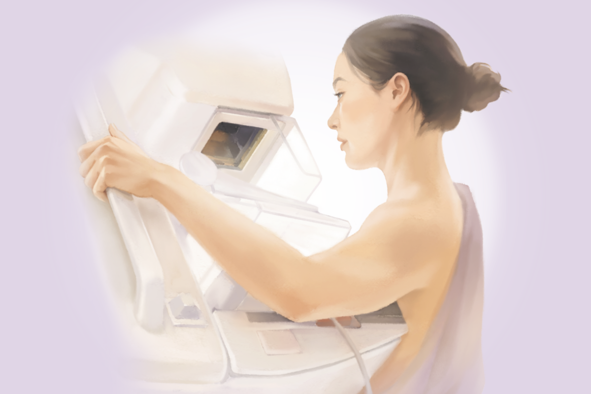
 (Illustration by The Epoch Times, Shutterstock)
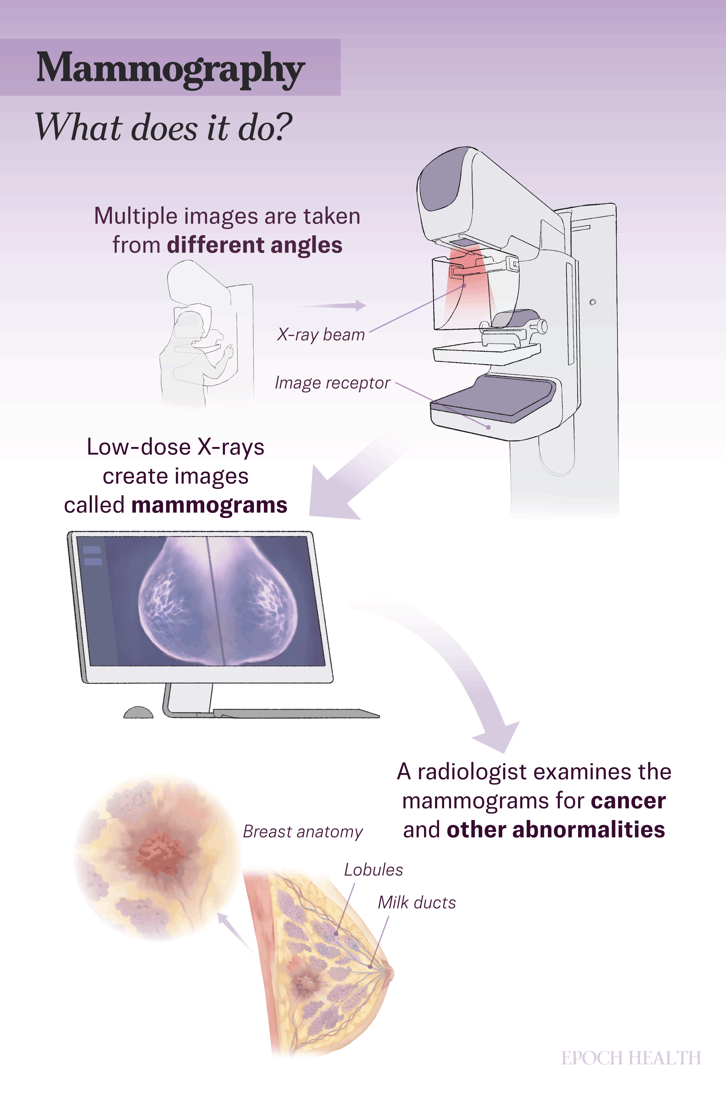
[shortcut_anchor id=”anchor_1781378642032″ label=”How It Works”]What Does Mammography Do?[/shortcut_anchor]
Mammography is an imaging test that uses low-dose X-rays (though higher than dental X-rays) to create images, called mammograms, of the breast. During the exam, each breast is placed between two plastic plates and gently compressed to flatten the tissue. Multiple images are taken from different angles, with compression lasting about 10 to 15 seconds per image. The whole exam typically takes about 20 minutes.
Mammography is commonly used in two ways. Screening mammograms can detect tumors at an early stage, often before symptoms appear. Diagnostic mammograms are used to evaluate breast changes, such as a lump or other abnormalities. Both use the same equipment, although diagnostic mammograms require additional images from multiple angles, resulting in a slightly higher radiation dose.
Three-dimensional mammography, also known as breast tomosynthesis, may be used alongside standard mammography. Unlike traditional mammography, which captures a few flat, two-dimensional images from fixed angles, breast tomosynthesis uses an X-ray arm that moves in an arc around the breast to obtain multiple images from different angles and create a clearer view of the breast.
This improved detail helps doctors distinguish normal overlapping tissue from potential cancer, leading to as much as a 40 percent increase in early cancer detection and a 40 percent reduction in false alarms. It’s particularly useful to women with dense breast tissue, although it involves more radiation and may carry additional costs not covered by all insurance plans.
[shortcut_anchor id=”anchor_1781378658532″ label=”Who Needs It”]Who Should Consider Mammography?[/shortcut_anchor]
Mammography screening guidelines vary depending on a woman’s risk level and the medical organization issuing the recommendations.
The U.S. Preventive Services Task Force recommends a mammogram every two years for women ages 40 to 74 who are at average risk. The American Cancer Society recommends annual mammograms for women aged 45 to 54, then every two years from age 55 onward.
Women at higher risk for breast cancer are advised to consult their doctor about beginning mammograms before age 40, screening more frequently, or adding other imaging tests such as ultrasounds or MRI. Higher-risk factors include a BRCA1 or BRCA2 gene mutation, a personal or family history of breast or ovarian cancer, dense breast tissue, or prior radiation therapy to the chest.
Even though the test has helped many women to detect breast cancer early and lower their risk of death, there has been discussion on whether every woman should undergo the screening to get the same benefits.
In recent years, concerns about overdiagnosis and overtreatment have gained increasing attention.
Overdiagnosis occurs when screening detects cancers that would not have caused symptoms or health problems during a person’s lifetime. These cancers may grow very slowly, remain stable, or might even regress on their own. A 2023 meta-analysis of 24 studies found that among women aged 40 to 89, about 12.6 percent of breast cancers detected through mammograms may fall into this category. Overdiagnosis rates also rise with age. A 2023 study of more than 54,000 women found that 31 percent of women aged 70 to 74, 47 percent aged 75 to 84, and 54 percent of women aged 85 and older were overdiagnosed.
Overtreatment follows directly from overdiagnosis. Because it is not currently possible to determine which early-stage breast cancers will progress and which won’t, most of them are typically treated. That means some people undergo surgery, radiation, or other therapies without any meaningful benefit while still facing physical and emotional cost.
A few special situations are worth noting:
- Women Aged 75 and Older: Screening decisions are typically individualized rather than based on routine screening schedules. This is because many breast cancers in this age group grow more slowly, and other health conditions often take precedence. However, cancers that are not found early may be diagnosed later at a more advanced stage, which can limit treatment options in some cases.
- Pregnancy and Breastfeeding: Mammograms are considered generally safe during pregnancy and breastfeeding, but are often postponed during pregnancy unless there are specific concerns.
- Breast Implants: Women with breast implants can still get mammograms. Informing the mammography facility ahead of time allows technologists to use special imaging techniques that move the implant aside for a better view. Women with reconstructed breasts after mastectomy typically don’t need screening on the reconstructed side.
- Men at Increased Genetic Risk: Some men have an inherited genetic risk for breast cancer, including BRCA1 or BRCA2 gene mutations. Routine screening mammograms are not recommended, but diagnostic mammograms may be used if symptoms appear.
[shortcut_anchor id=”anchor_1781378672538″ label=”Effectiveness”]How Effective Is Mammography?[/shortcut_anchor]
Mammography identifies about 87 percent of breast cancers, with higher accuracy in older women and those with fatty, or less dense, breasts.
The survival benefits of screening mammography vary by age. Research shows that mammography is associated with about a 14 percent reduction in breast cancer mortality among women in their 50s, and about a 33 percent reduction among women in their 60s.
For women in their 40s, the benefit exists but is smaller and less statistically clear. However, more recent studies offer a more encouraging view for earlier screening. A 2022 study and a 2024 study both found that annual mammograms starting at age 40 and continuing into the late 70s or beyond were associated with up to a 40 percent reduction in breast cancer deaths.
Mammography is also effective in detecting many types of breast cancer, including invasive ductal carcinoma and invasive lobular carcinoma, as well as small growths such as ductal carcinoma in situ—an early-stage abnormal tissue growth confined to the milk ducts that hasn’t yet spread into surrounding tissue.
However, there may still be false positives and false negatives.
About 10 percent to 12 percent of mammograms in women aged 40 to 49 result in a false alarm. Over 10 years of annual screening, about half of women will experience at least one false-positive result, and 7 percent to 12 percent will receive at least one recommendation for a biopsy that ultimately does not find cancer. The emotional effect of a false alarm can also linger for years, with some women continuing to experience symptoms of stress and depression long after being declared cancer-free.
A false-negative result occurs when a mammogram misses a cancer that’s actually there. About 1 in 8 breast cancers goes undetected by screening mammography, most often in women with dense breast tissue, where tumors can be harder to distinguish from surrounding tissue. This is why it is important to consult a doctor if any new breast symptoms appear, even after a recent normal mammogram.
Screening also doesn’t seem to significantly improve all-cause mortality rates.
[shortcut_anchor id=”anchor_1781378684748″ label=”Risks”]What Are the Risks of Mammography?[/shortcut_anchor]
Although mammography is generally considered safe, it still has risks, including:
- Radiation Exposure: Radiation exposure from a single mammogram is low—roughly equivalent to the natural background radiation a person absorbs over about five weeks. However, the breast is one of the body’s more radiation-sensitive organs, and some researchers suggest that cumulative exposure over decades of annual screening may be worth considering as part of an individual risk assessment.
- Discomfort: Mammography is generally well tolerated with few complications. Possible side effects from breast compression include temporary discomfort, bruising, or small hematomas. Inadequate images can usually be prevented with proper positioning and technique.
- Anxiety: Mammography can cause anxiety among women before the exam, while waiting for results, or after receiving an abnormal finding that requires additional testing. False-positive results can further increase stress and emotional distress, even when no cancer is ultimately diagnosed.
[shortcut_anchor id=”anchor_1781378697931″ label=”Alternatives”]What Are the Alternatives to Mammography?[/shortcut_anchor]
Mammography remains the preferred method for breast cancer screening. Growing awareness of the limitations of mammography, especially in women with dense breast tissue, has led more people to explore supplemental and alternative screening options and approaches beyond a standard mammogram.
1. Breast MRI
This highly sensitive imaging test uses magnets and radio waves to create detailed images of the breast. It is mainly used alongside mammography for women at high risk of breast cancer, such as those with BRCA mutations.
Breast MRI is more sensitive than mammography and can detect some cancers that mammograms may miss, especially in women with dense breast tissue or those at high risk. However, it is more expensive, takes longer to perform, and is more likely to produce false-positive results.
2. Breast Ultrasound
Breast ultrasound is primarily used as a follow-up imaging test used to clarify abnormalities found on a mammogram or physical exam, helping distinguish between fluid-filled cysts and solid masses.
It’s also useful in evaluating women with very dense breast tissue and during pregnancy because it doesn’t use radiation. However, ultrasound isn’t a reliable primary screening method on its own, as it tends to miss small calcifications and other subtle findings that mammography detects, and it has a high false-positive rate.
3. Thermography
Thermography, also known as digital infrared thermal imaging, uses an infrared camera to detect heat patterns on the skin surface that may signal inflammation or early physiological changes before a structural abnormality is detectable. It is noninvasive, painless, and does not use radiation or compression, making it safe for repeated scans.
Annual thermography scans can establish a personalized baseline, allowing changes in a patient’s heat patterns over time to help identify potential problems early. However, evidence supporting thermography as a standalone breast cancer screening test remains limited. Results can also be skewed by factors such as exercise, caffeine, certain medications, and room temperature. It is best considered as a complementary tool rather than a replacement for mammography.
4. Molecular Breast Imaging
Molecular breast imaging uses a small radioactive tracer to highlight cancer cells that may be hidden on mammograms due to dense breast tissue. Because it detects tissue activity rather than just structure, it can identify some cancers that mammography misses.
However, it uses more radiation, is less widely available, takes longer, and costs more than mammography, so it is mainly used as a supplemental test.
A 2025 study involving nearly 3,000 women followed over several years found that adding molecular breast imaging to tomosynthesis detected about 2.5 times more invasive breast cancers than mammography alone, although it also slightly increased the number of women called back for additional testing.
5. Contrast-Enhanced Mammography
This imaging technique uses injected iodine contrast and specialized mammography to highlight areas of increased blood supply in the breast, which can help identify tumors. It is considered a lower-cost, more accessible alternative to breast MRI and can approach the accuracy of MRI for staging breast cancer and monitoring treatment response. However, the contrast injection carries a small risk of allergic or kidney-related complications.
[shortcut_anchor id=”anchor_1781378734749″ label=”Preparation”]How Do I Prepare for a Mammography?[/shortcut_anchor]
Before scheduling a mammogram, you should discuss any new breast changes, prior surgeries, hormone use, and personal or family history of breast cancer with your doctor. It is generally best to avoid scheduling the exam during the week before menstruation, when the breasts are more tender. The ideal timing is often one week after your menstrual period. If you have had mammograms at another facility, obtain copies so the radiologist can compare them with your current images.
On the day of the mammogram:
- Avoid applying deodorant, lotions, powders, perfumes, or similar products under the arms or on the breasts, as they can interfere with image quality.
- Wear clothing that is easy to remove and avoid jewelry around the neck or chest.
- Consider taking a mild pain reliever about an hour before the appointment, after consulting your doctor, to reduce discomfort.
Some state and local programs, including the Centers for Disease Control and Prevention’s National Breast and Cervical Cancer Early Detection Program, provide free or low-cost mammograms and related screening services for low-income or uninsured women.
[shortcut_anchor id=”anchor_1781378753165″ label=”Recovery”]What to Expect After a Mammography?[/shortcut_anchor]
After a mammography, two radiologists—or one radiologist assisted by artificial intelligence—review the images. Results from a screening mammogram are typically available within two weeks. Don’t assume your results are normal if you don’t hear back. It’s worth following up if you haven’t received your report.
Your results will include a BI-RADS score, a standardized rating system that guides follow-up care. Scores range from negative or benign findings—which require routine screening—to probably benign findings—which typically require a six-month follow-up scan—to suspicious or highly suggestive results that may require a biopsy.
The report will also note your breast density. Dense breast tissue is common and can only be detected through mammography. Breast density matters because it slightly increases breast cancer risk and makes mammograms more difficult to interpret.